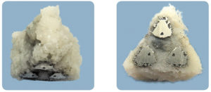
Credit: PRNewsphoto / SI-Bone, Inc.
The popularity of sacroiliac joint fusion has grown rapidly over the past five years, with SI-Bone’s Ifuse device leading the market. Recently approved by the US Food and Drug Administration, the company has released a new version of their traditional triangular titanium implants. The second-generation products (Ifuse-3D implants) are made using additive manufacturing, and incorporate a fenestrated design and porous surface. According to new research published in the International Journal of Spine Surgery, the new Ifuse-3D implant resulted in significantly greater bone infiltration ingrowth and through growth than the original Ifuse device.
Regina MacBarb, (San Jose, USA) and colleagues used a bilateral ovine distal femoral defect model to compare the Ifuse device—machined solid triangular titanium plasma spray-coated sacroiliac joint implants—and the new Ifuse-3D device, used independently, with nanocrystalline hydroxyapatite (HA) or pre-packed with autograft.
Bone growth in all implants
Radiographic, biomechanical and histomorphometric evaluations took place at six and twelve weeks post-implantation of two implants into each of 24 female Suffolk sheep. The four test groups were as follows: Ifuse, Ifuse-3D, Ifuse-3D+HA and Ifuse-3D+autograft.
Peri-implant bone was observed on radiographic results for all implants, with continuous formation of bone through the Ifuse-3D implants. These microcomputed tomography results were confirmed through histological analysis. Pushout testing results did not differ significantly between the four groups at six or twelve weeks; however, gross examination of post-pushout testing samples revealed “spot-welds” of bone remaining attached to the Ifuse implants, whereas a ~2–3mm continuous ring of integrated bone remained attached to all Ifuse-3D implant groups following testing.

All groups demonstrated some bone maturation (50:50, lamellar to woven bone) at six weeks on semi-quantitative histomorphetric analysis, while both the Ifuse and the Ifuse-3D+autograft group reached 50–75% lamellar bone at 12 weeks. Within the Ifuse-3D implants themselves, both the Ifuse-3D and Ifuse-3D+autograft groups demonstrated 50% lamellar bone at 12 weeks, while the bone maturation through the centre of the Ifuse-3D+HA group did not progress beyond 25% lamellar bone during the experiment.
In terms of porous surface, the Ifuse-3D implants offered 1.4 times the length and 2.8 times the surface area of the original Ifuse implants. This was mirrored by a significantly greater bone ingrowth at six and 12 weeks in all Ifuse-3D groups. By 12 weeks, the Ifuse-3D+autograft implants performed better still, with significantly more bone ingrowth than both the Ifuse (p<0.001) and the Ifuse-3D+HA (p<0.01) groups.
Adding together the amount of bone filling the fenestrated core as well as the porous surfaces of the Ifuse-3D implants at 12 weeks, the researchers found them to have in total 2.5 times (Ifuse-3D), 2 times (Ifuse-3D+HA) and 3.3 times (Ifuse-3D+autograft) more bone fill than the Ifuse group.
Conclusions
The Ifuse-3D implants “all experienced significantly more bony ongrowth, ingrowth and through growth” compared to the Ifuse group, the authors reported. While both implants “formed stable interfaces with host bone, these results indicate that they each have distinct mechanisms for promoting implant stabilisation that may influence the degree of biological fixation.”
The authors note that, “the differing push-out failure mechanisms encountered in the present study may demonstrate that the integrity of the bone integration” of the Ifuse-3D implants exceeds that of the Ifuse implants.
Concluding, they write that the Ifuse-3D implants offer “a highly interconnected porous surface with an open fenestrated core structure that offers comparatively greater surface area for bony integration and the ability to be pre-packed with graft material”. In addition, whilst the nanocrystalline HA did not benefit fusion, the combination of the new implant with autograft provided significantly improved results.
The research was published in June 2017 in the International Journey of Spine Surgery. doi: 10.14444/4016.
This article was originally published in Spinal News International 44.


