
In a market booming with new technologies and therapies, spinal surgeons are spoilt for choice when it comes to implants and other devices. Stryker’s John Mayor talks to Spinal News International about emerging technologies, education and the evidence behind the hype.
What do you think are the greatest challenges facing spinal surgeons today?
As technology continues to change rapidly, it can be a challenge for spinal surgeons to keep up with the latest advances and determine which technologies best meet their needs and those of their patients.
For example, a growing number of companies are using 3D-printing (also known as additive manufacturing) technologies to create spinal implants. Not all 3D-printed implants are the same, however. Some offer porous surface technology while others use a fixed lattice pattern. At Stryker, we use a randomised porous structure mimicking cancellous bone. It is important for spine surgeons to understand these differentiators.
How can physicians keep up-to-date with emerging technologies?
Education is key. We have worked hard to educate surgeons about our 3D-printed implants.
This includes inviting surgeons to Stryker’s manufacturing facility to see first-hand how the company’s Tritanium in-growth technology enables its spinal implants to be designed and built with pinpoint precision, and to optimise pore size, porosity, geometry, and surface texture.1

Stryker has invested significantly in 3D printing. Why do you expect this manufacturing method to be so successful?
Additive manufacturing technology has the ability and flexibility to create geometries previously difficult or impossible to manufacture, produce minimal waste, and potentially offer improved product development speed.
While additive manufacturing may appear to be a recent development in the creation of medical implants, momentum has actually been building for some years. Stryker began collaborations with aerospace companies and top universities in the UK and Ireland in 2001 to industrialise 3D printing for the healthcare industry.
Tritanium, the company’s novel, highly porous titanium alloy material, is made using a proprietary additive manufacturing process. The Tritanium PL cage is built layer by layer, which enables the creation of complementing porous and solid structures in a single device. These structures are designed to resemble cancellous bone.1,2
The material is the culmination of nearly 15 years of extensive research, development, and validation in material science and manufacturing, and it has been utilised clinically for more than 10 years—with more than 300,000 knee and hip devices implanted.
Today, Stryker makes and sells more metal implants using additive manufacturing than any company in the world and anticipates growth of its Tritanium PL cage to double in 2017. The next spinal implant in the product line will be a cervical cage.

The importance of porosity is becoming increasingly clear for fusion. How porous are Tritanium cages?
The Tritanium PL cage is created with fully interconnected pores that span endplate to endplate, with a mean porosity of 55-65% and a mean pore size of 400-500μm. The porous matrix of the cage is engineered by computer models based on studies investigating which geometry and pore size may provide an optimal environment for cells to attach and multiply within this structure.2,3 Its roughened porous surface is designed to enhance interconnectivity and provide a stronger interface with surrounding bone compared to a smooth surface.4,5
This “precise randomisation”2 of fully interconnected pores contrasts with other spinal implants that have longitudinal channels and traverse windows that result in a uniform lattice structure, as well as cages offering porosity that is only present on the surface.

How has Tritanium been investigated scientifically?
A pre-clinical animal study was conducted to compare the biomechanical, radiographic, and histological performance of spinal implants with different surface technologies in an ovine lumbar interbody fusion model.
The interbody fusion cages involved in this study included traditional PEEK cages, plasma-sprayed titanium-coated PEEK cages, and Stryker’s 3D-printed Tritanium PL cages.
Results of the study were presented at the North American Spine Society’s annual meeting (NASS; 26–29 October 2016, Boston, USA) and at the American Academy of Orthopaedic Surgeons annual meeting (AAOS; 14–18 March, San Diego, USA).
A recent wicking study demonstrated that a cube built with Tritanium material was able to wick fluid into the porous structure under specific conditions, and it also absorbed and held fluid inside the porous structure.7 This experiment was performed using heparinised porcine bone marrow aspirate.
An 18-day cell attachment study is also being conducted to evaluate how Tritanium in-growth technology may create an optimal environment for cell attachment
In addition to this, The Bone and Joint Clinic of Baton Rouge, Baton Rouge, USA, is conducting a clinical study to evaluate the fusion rate and clinical outcomes of patients with degenerative disc disease who undergo a transforaminal lumbar interbody fusion (TLIF) with a Tritanium cage.6
Stryker has had a number of other product releases over the past year, including the LITE Bio delivery system, the Aero-C ACDF device and the Xia cortical trajectory system platform. What is innovative about these products?
LITE BIO delivery system:
The LITE BIO delivery system is designed to provide surgeons with a single-handed method to deliver autograft, allograft, or synthetic bone graft material without obstructing visibility. The innovative delivery tool provides tactile and visual confirmation of bone graft delivery, and the mallet-free system eliminates the impaction of bone graft. The low-profile design allows visibility through a decompression tube without obstructing view. A radiolucent strip facilitates visualisation under fluoroscopy, and the disposable cannula allows for delivery of up to 5cc of bone graft at one time.
The system was recently named as a finalist for the Technology Innovation Award as a part of Back Pain Centers of America’s annual awards series.
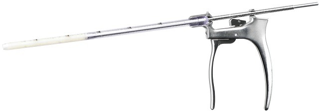
Aero-C:
![]()
Aero-C is the only straightforward anterior cervical discectomy and fusion (ACDF) device intended to offer uniform compression across the interbody space. Using Aerofoil compression technology, Aero-C is designed to pull the vertebral body toward the implant as it is inserted, creating compressive forces at the implant-to-endplate interface.8 Aerofoil compression technology is also available for lateral and anterior lumbar interbody fusion procedures (LLIF and ALIF). Since the initial introduction to the market, nearly 900 cases have been completed.
Xia CT
 Xia CT implants and instruments are used in “less invasive” LITe LIF posterior lumbar interbody fusion procedures for patients with degenerative disc disease, spondylolisthesis and trauma. The cortical trajectory procedure facilitates a smaller midline incision to help achieve decompression, fixation, and fusion.9 It also is intended to be more muscle sparing than standard open procedures that require lateral dissection, and its reduced incision may allow for more efficient exposure and closure time.9 The Xia CT system was introduced in 2016, and more than 915 cases have been completed to date.
John Mayor is vice president of Global Marketing at Stryker.
References
- PROJ*43909: Tritanium Technology Claim Support memo.
- Karageorgiou V, Kaplan D. Porosity of 3D biomaterial scaffolds and osteogenesis. Biomaterials, 2005. 26: 5474-5491.
- ID TRITA-LM-2: 3D-Printed Tritanium—Advancing Spinal Surgery
- McGilvray KC, et al. Biomechanical and histologic comparison of a novel 3-D printed porous titanium interbody cage to peek. The Spine Journal, 2016. 16(suppl. 10): S363–364
- Deligianni DD, Katsala N, Ladas S et al. Effect of surface roughness of the titanium alloy Ti-6Al-4V on human bone marrow cell respose and on protein adsorption. Biomaterials. 2001;22(11):1241-51.
- “Clinical and Radiological Evaluation of Patients With DDD Following TLIF With 3-D Printed Titanium Cage”gov. Web. 05 June 2017. https://clinicaltrials.gov/ct2/show/NCT03018392
- RD0000050927
- PROJ0000050417 Aero-C Anchor Induced Compression Testing Design Iteration Memo
- Lee GW, Son JH, Ahn MW, Kim HJ, Yeom JS. (2015) The comparison of pedicle screw and cortical screw in posterior lumbar interbody fusion: a prospective randomized noninferiority trial. The Spine Journal 15, 1519-1526.


